
How to Choose the Best Suture Anchor for Ankle Repair?
Choosing the best suture anchor for ankle repair requires more than comparing product sizes or prices. The surgeon must match the anchor to the injury, tissue quality, fixation site, and planned rehabilitation. A strong Suture Anchor Ankle solution should support stable fixation without creating unnecessary bone damage.
In clinical practice, small differences matter. A lateral ligament repair may involve thin, delicate tissue near the fibula. An anchor with suitable diameter, insertion depth, and pullout resistance can improve handling during fixation. Material also deserves careful attention. Bioabsorbable, all-suture, and metallic anchors each offer different benefits and limitations. The final choice should follow current evidence, manufacturer instructions, and the surgeon’s experience with ankle anatomy.
No anchor is perfect. That matters.
This guide examines the practical factors behind anchor selection, including load requirements, knot management, imaging considerations, and compatibility with minimally invasive techniques. It also considers patient-specific issues, such as bone density, athletic demands, previous surgery, and the risk of delayed healing. Laboratory strength does not always predict clinical success. That assumption can fail.
Reliable decisions come from combining peer-reviewed research, verified technical data, and careful intraoperative judgment. Surgeons should also review local protocols and discuss uncertain cases with qualified colleagues. The goal is not to promote one design. It is to clarify how each feature may influence safety, efficiency, and long-term ankle stability. With a structured comparison, clinicians can make better-informed choices for each repair rather than relying on habit alone.
Understanding Suture Anchors in Ankle Repair
How to Choose the Best Suture Anchor for Ankle Repair?
Understanding Suture Anchors in Ankle Repair
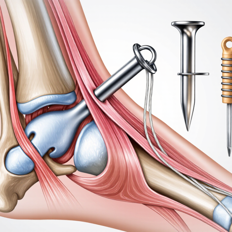
Suture anchors secure damaged ligaments or tendons to bone during ankle repair. Their performance depends on more than size or shape. The surgeon must assess bone quality, tear location, tissue strength, and required fixation depth. A small anchor is not always safer. Weak bone may need a different design or placement strategy. The insertion angle also matters, especially near narrow ankle structures. Poor alignment can reduce fixation and irritate nearby tissue.
Tips: Match the anchor to the repair, not the other way around. Check thread strength, anchor stability, and compatibility with the surgical technique. Confirm the device suits the patient’s bone condition. Careful measurement helps prevent excessive drilling. Small details matter.
Material selection deserves equal attention. Some anchors are absorbable, while others remain in the bone. Each option may offer advantages and limitations. The choice should consider healing expectations, imaging needs, and the surgeon’s familiarity with the system. In clinical practice, reliable handling can be as important as laboratory strength. A technically strong anchor may still perform poorly if insertion is rushed. That is worth reflecting on. Surgeons should review manufacturer instructions, current evidence, and patient-specific risks before choosing. There is no universal best anchor. The most appropriate option supports stable fixation with minimal tissue disruption.

Identifying the Ankle Injury and Repair Requirements
Choosing the best suture anchor begins with identifying the ankle injury, not selecting a device. An ankle is not a single structure. A lateral sprain may involve the anterior talofibular ligament, calcaneofibular ligament, capsule, or syndesmosis. The National Athletic Trainers’ Association reports that ankle sprains represent approximately 10–30% of sports injuries. That range matters. It shows why a standard repair plan can be unreliable.
Clinical examination should locate tenderness, instability, swelling, and loss of function. The Ottawa Ankle Rules help screen for fractures, while the American College of Radiology recommends imaging based on trauma findings and persistent symptoms. Magnetic resonance imaging can clarify ligament quality, cartilage damage, or an occult fracture. Ultrasound may show dynamic ligament gapping, but operator skill changes its reliability. The 2021 Journal of Orthopaedic & Sports Physical Therapy guideline also stresses evaluating syndesmotic injury and recurrent instability. Do not treat the scan alone.
Repair requirements then become more specific. Poor tissue may need reinforcement, while good ligament remnants may support a direct repair. Bone density, footprint size, loading demands, and the number of fixation points influence anchor selection. A small footprint may require careful tension control. Excessive tension can restrict motion. In practice, this step is often rushed. A neat anchor choice can still fail when the diagnosis is incomplete. The surgeon should document injury grade, tissue quality, bone condition, and rehabilitation demands before final fixation planning.
Comparing Suture Anchor Materials, Designs, and Fixation Strength
Choosing an ankle suture anchor requires more than comparing maximum pullout strength. Material matters. Nonabsorbable polymers provide durable support, while bioresorbable materials may reduce long-term implant presence. However, degradation can affect local tissue response and strength over time. Metal anchors can offer high initial stability, but they may complicate revision procedures or create imaging concerns.
Design also changes performance. Screw-in anchors can provide strong purchase in dense bone. All-suture anchors use a smaller drill hole and may preserve bone stock. Knotless systems reduce knot prominence, while knotted anchors allow tension adjustment during repair. Fixation strength depends on anchor geometry, insertion angle, tendon quality, and bone density. A strong anchor in weak bone may still pull out. It happens.
Tips: Match the anchor to the lesion and bone. Check cyclic loading data, not only ultimate strength. Confirm the drill depth and insertion angle. Leave enough tendon tissue for secure tensioning. When evidence is limited, document the reasoning and avoid assuming that newer designs are automatically better.
Surgeons should evaluate laboratory results alongside clinical outcomes and handling experience. Cadaver testing may not reflect swollen, osteoporotic, or repeatedly loaded ankles. That gap deserves attention. A reliable choice balances material behavior, design efficiency, fixation strength, and the patient’s rehabilitation demands. No single anchor suits every repair.
Selecting the Right Anchor for Bone Quality and Surgical Technique
How to Choose the Best Suture Anchor for Ankle Repair?
Selecting the right suture anchor begins with bone quality, not convenience. Dense cortical bone may hold a smaller anchor securely. Soft or osteoporotic bone requires greater attention to thread design, diameter, and insertion depth. A preoperative scan can reveal cysts, fractures, or reduced bone stock around the repair site. Plain radiographs may not show every concern. That matters.
Surgical technique should guide the anchor choice. For lateral ankle ligament repair, the surgeon must match anchor placement with the ligament’s native footprint. A perpendicular insertion angle can improve purchase and reduce pullout forces. In a tight working space, a low-profile delivery system may offer better control. Keep the drill channel clean. Excessive tapping can weaken fragile bone. Tension the suture gradually, while checking tissue contact and ankle motion.
Material selection also deserves careful judgment. Nonabsorbable sutures can provide lasting support, while absorbable options may reduce long-term foreign material. Each choice has trade-offs. Clinical evidence, patient activity, bone density, and revision plans should shape the decision. I would not select an anchor from size alone. Small anchors can fail in poor bone, yet oversized anchors may damage a narrow insertion site. Even experienced surgeons should reassess the plan when intraoperative bone quality differs from imaging. A secure repair is not simply tight; it is anatomically placed, stable, and compatible with rehabilitation.
How to Choose the Best Suture Anchor for Ankle Repair?
Selecting the Right Anchor for Bone Quality and Surgical Technique
Material stiffness provides context, but it does not independently determine pullout strength or clinical success. In poor cancellous bone, fixation is influenced by thread design, anchor diameter, insertion depth, cortical purchase, footprint preparation, and load-sharing technique. Metallic anchors generally provide high stiffness, while polymer and bioresorbable anchors may reduce imaging artifact and allow different biological or revision considerations.
Values are approximate elastic-modulus ranges reported for commonly used implant materials and bone types; values vary with composition, porosity, hydration, testing method, and patient-specific bone quality. Final anchor selection should follow the device instructions and the surgeon’s intraoperative assessment.
Evaluating Placement, Complications, and Postoperative Considerations
Choosing the best suture anchor for ankle repair requires more than comparing pullout strength. Placement, tissue quality, and postoperative demands matter equally. A 2022 systematic review in Foot & Ankle Orthopaedics reported that complication rates after ankle ligament surgery varied widely, partly because surgical techniques and definitions differed. This inconsistency deserves attention.
For lateral ligament repair, the anchor should sit in the intended ligament footprint, not merely in convenient bone. Surgeons commonly use imaging to confirm tunnel direction and avoid the joint surface, cartilage, and nearby tendons. Poor trajectory may cause cortical breakout or anchor prominence. Small anchors can reduce bone removal, but they may provide less margin in osteoporotic bone. Bigger is not automatically safer. The best choice depends on footprint size, bone density, and load requirements.
Postoperative planning should begin before insertion. A 2023 review in the Journal of ISAKOS linked early rehabilitation decisions with repair protection, swelling control, and patient-specific risk factors. Protected weight bearing may be necessary when fixation is weak or tissue quality is poor. Wound irritation, nerve symptoms, infection, stiffness, and recurrent instability require active surveillance. The American Orthopaedic Foot & Ankle Society emphasizes patient-reported function and stability, not imaging alone, when evaluating recovery. That is practical, but imperfect. Imaging can look reassuring while pain persists. Conversely, mild radiographic changes may have little clinical meaning. Documenting anchor position, fixation quality, and rehabilitation milestones improves communication and supports safer postoperative decisions.
| Repair Scenario | Common Anchor Configuration | Key Placement Considerations | Primary Benefits | Important Risks and Complications | Postoperative Priorities | Selection Factors |
|---|---|---|---|---|---|---|
| Lateral ankle ligament repair | One or more small-diameter suture anchors placed at the ligament’s anatomical insertion. Knotless or knotted constructs may be used according to tissue quality and surgeon preference. |
|
|
|
| High priority: anatomical accuracy, adequate bone stock, soft-tissue quality, and construct strength. |
| Medial ankle ligament repair | Suture-anchor repair or augmentation of the deltoid ligament, often using multiple fixation points when the injury involves more than one ligament layer. |
|
|
|
| High priority: injury pattern, ligament-layer involvement, bone quality, and avoidance of excessive tension. |
| Syndesmotic stabilization | Suture-button or screw-based fixation may be selected; a suture anchor alone is generally not the standard construct for an unstable syndesmosis. |
|
|
|
| Do not substitute automatically: choose fixation based on demonstrated syndesmotic instability rather than the availability of an anchor. |
| Osteochondral lesion or cartilage-related fixation | Small, low-profile fixation may be considered only for selected fragment or soft-tissue stabilization procedures; anchor suitability depends on lesion location and bone depth. |
|
|
|
| Very high priority: low profile, controlled insertion depth, and precise imaging or arthroscopic guidance. |
| Tendon or retinacular repair | Small suture anchors or transosseous sutures may be used for peroneal retinaculum, Achilles-region, or other tendon-related repairs, depending on anatomy and tissue loading. |
|
|
|
| High priority: tendon load, tissue thickness, anchor prominence, and rehabilitation compliance. |
| Revision repair or poor bone quality | Larger or alternative fixation may be necessary; options can include multiple anchors, bone tunnels, graft augmentation, or reconstruction rather than a single anchor. |
|
|
|
| Highest planning priority: bone stock, prior surgery, tissue quality, patient risk factors, and the need for reconstruction. |
Article Source:
NORTH EASTON MACHINE • 218 Elm Street • North Easton, MA 02356 • 508-238-6219